Home
Research
Publications
Press
Outreach
Fun Stuff

Vaidehi Natu

Home
Research
Publications
Press
Outreach
Fun Stuff

Brain folds begin to form in the womb and deepen substantially throughout the first year of human life. In a new study we find that folds which emerge earlier in gestation—during roughly the 16th to 19th gestational weeks, such as the calcarine sulcus—are already relatively deep at birth and then show little to no additional deepening over the first postnatal year. By contrast, sulci that appear later in utero tend to deepen more rapidly after birth than those that formed earlier. This postnatal sulcal deepening reflects a complex, multicomponent process involving coordinated morphological changes and underlying microstructural development.

Tung et al., 2026 Nature Communications Biology

Want to learn how microstructural tissue properties develop in the visual cortex during the first year of human life? Our new study shows that the microstructure of the gray matter and adjacent white matter in brain regions across the dorsal, lateral, and ventral visual processing streams becomes denser after birth. This pattern indicates substantial tissue growth in the brain following an infant’s birth. Within each stream, early visual areas appear more mature at birth than higher-level areas, yet they show slower postnatal development compared with those higher-level areas. This hierarchical organization may be a core feature of the visual system, with newer processes building on and refining older ones. We hypothesize that if microstructural maturation is tied to functional specialization, then functional development may follow a similar hierarchical course.

Perez et al., 2026, Brain, Structure and Function

Screen Shot 2021-09-19 at 8.27.59 AM (2).png

In our exciting new study, using quantitative MRI and transcriptomic gene analysis, we show that babies in the first 6 months of life are extremely busy growing new gray matter tissue and cortical microstructures related to myelin and synapses in their primary sensory-motor systems. This tissue growth is hierarchical as lower-level areas are more developed at birth, but higher-level areas have faster rates of development after birth.

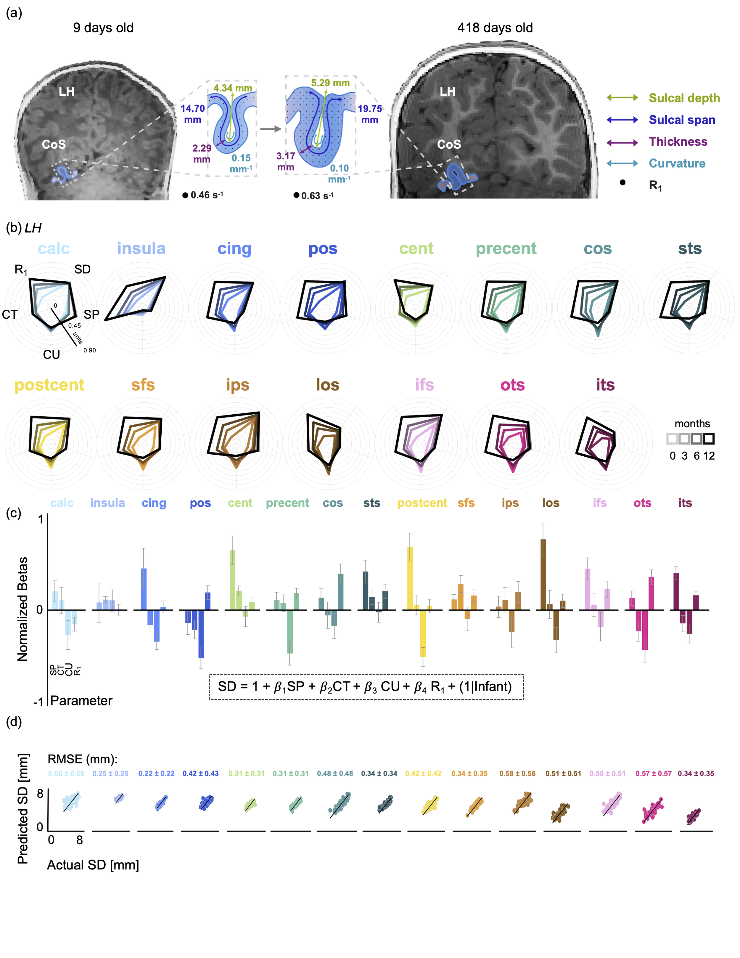
(Natu et al., 2021, Nature Communications Biology)

 
Screen Shot 2020-10-05 at 3.11.14 PM.png

There is a surprisingly well preserved structural-functional relationship in deep cortical folds of the high-level visual cortex across evolution and human development.

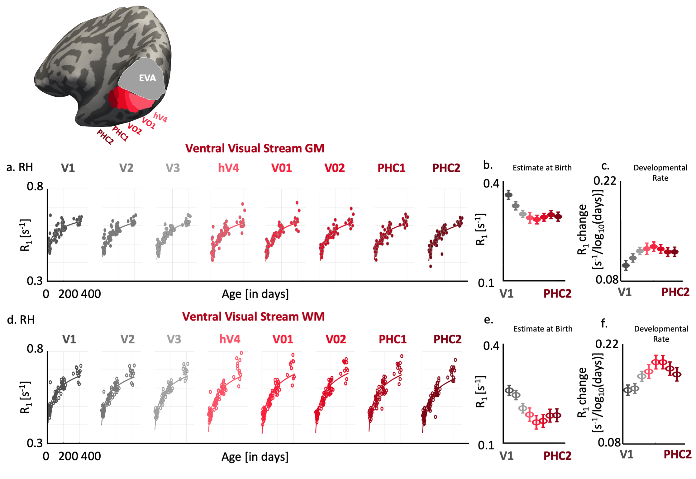
(Image from: Natu et al., 2020, Cerebral Cortex)

Screen Shot 2019-11-16 at 6.18.58 PM.png

Cortical tissue grows during child development in brain areas involved in reading and face memory. This t

issue growth is related to increase in myelination

(what is myelin? it is

a fatty tissue that insulates neural fibers and enables faster neural conduction) and dendritic arborization.

(Image from: Natu et al., 2019, PNAS, see also Gomez, Barnett, Natu et al., 2017, Science).

Screen Shot 2019-11-16 at 7.15.06 PM.png

I use

postmortem data, histological myelin/nissl stains to study the cellular and microstructural mechanisms underlying our MR findings.

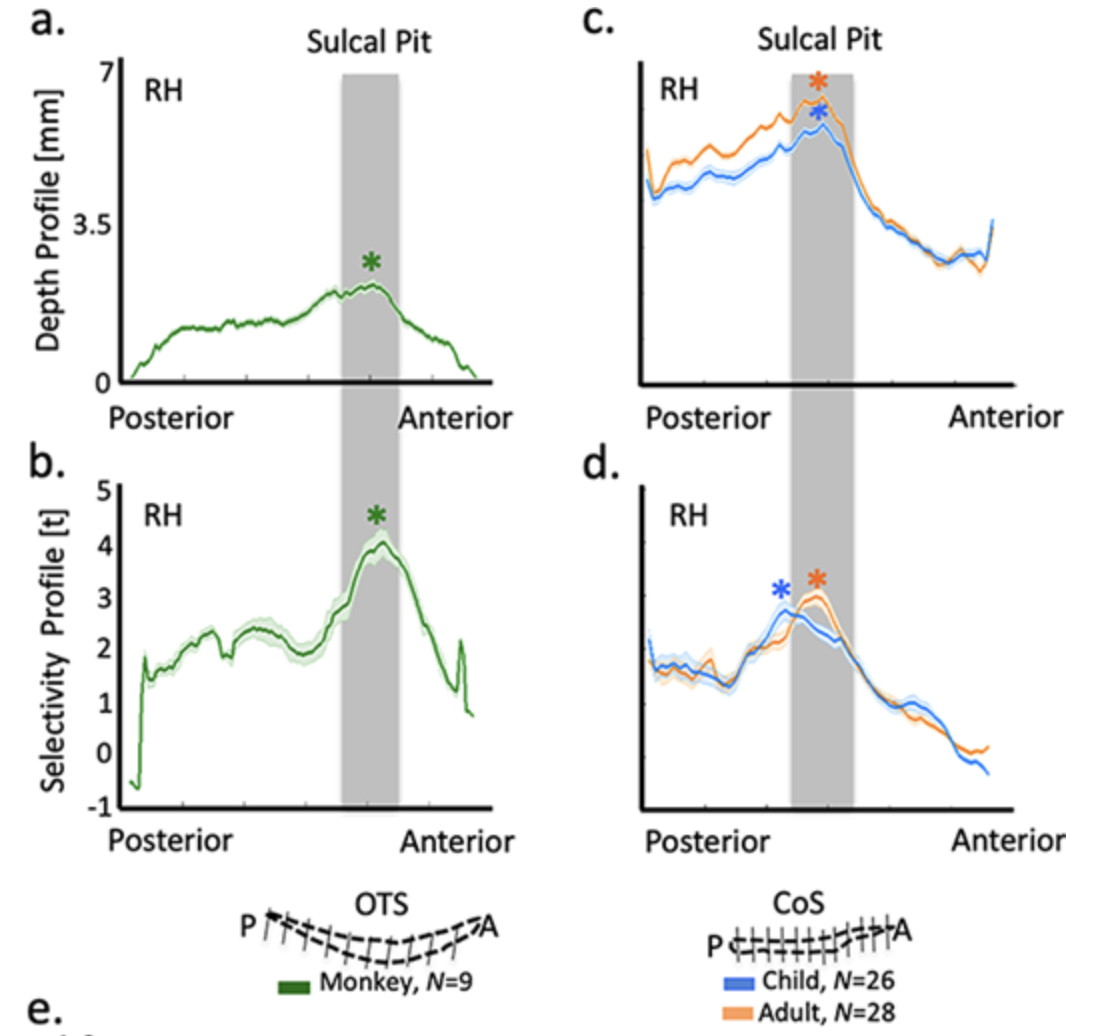
(Image from: Natu et al., 2019, PNAS).

Screen Shot 2019-11-16 at 6.43.33 PM.png

Receptive field properties of regions involved in face recognition and reading develop from childhood to adulthood by increasing the foveal coverage bias for faces in the right hemisphere and words in the left hemisphere.

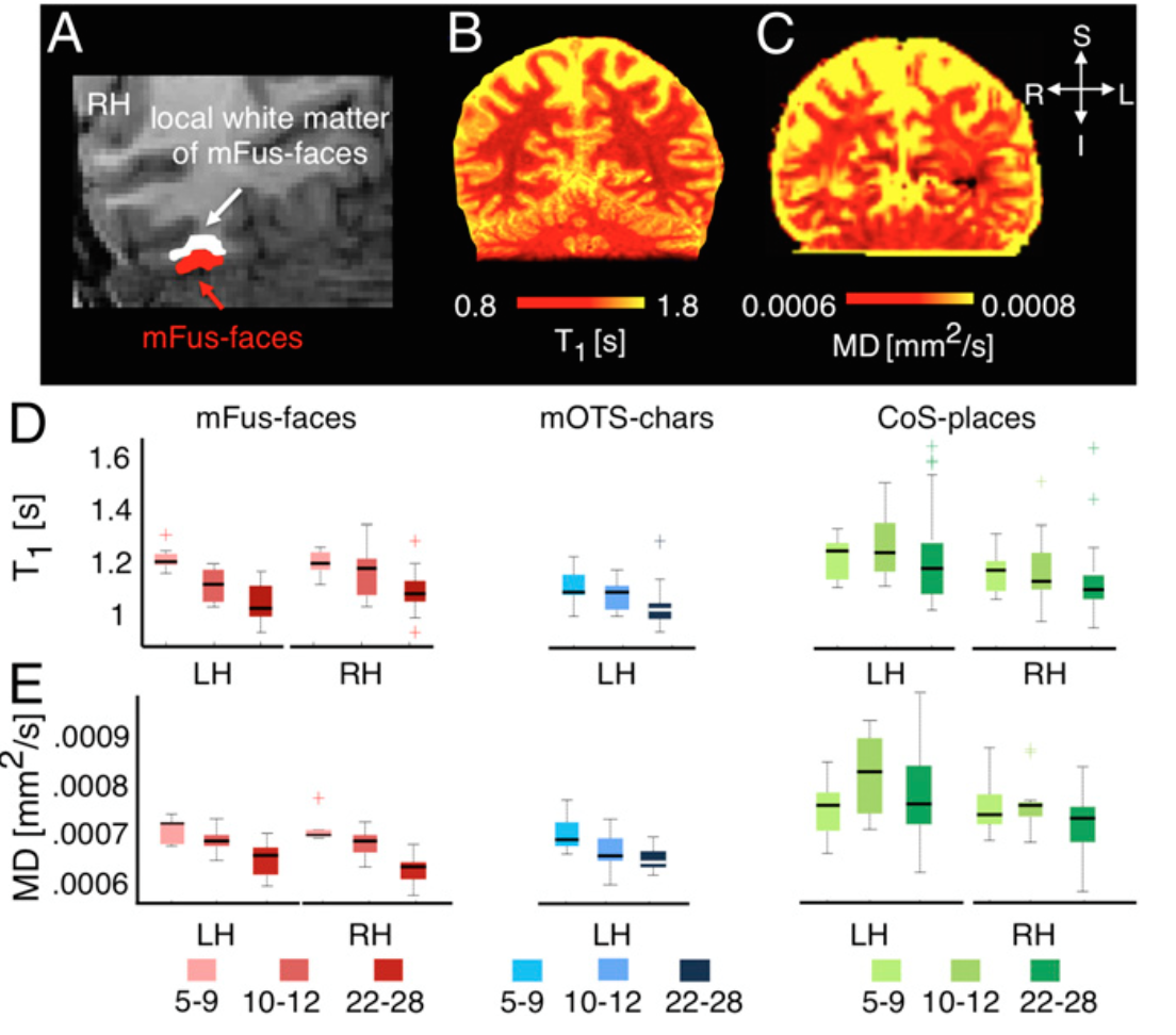
(Image from: Gomez, Natu et al., 2018, Nature Communications).

Screen Shot 2019-11-16 at 6.55.24 PM.png

Neural sensitivity to socially salient stimuli such as human faces increases with age in regions involved in face processing and is related to perceptual discriminability for faces. S

ensitivity is also dependent on our social milieu and experience, as neural sensitivity is stronger for own-age than other-age faces.

(Image from: Natu et al., 2016, Journal of Neuroscience).

a-The-neural-discriminability-of-the-brain-activity-patterns-elicited-in-response-to.png

Brain patterns are different for own-race faces than other-race faces.

Using pattern-based classification algorithms, my work showed that we can discriminate between brain’s response patterns for faces of one’s own race versus faces of a different race. This suggests that our social environment plays a critical role in shaping brain patterns as well as social biases for salient features such as race.

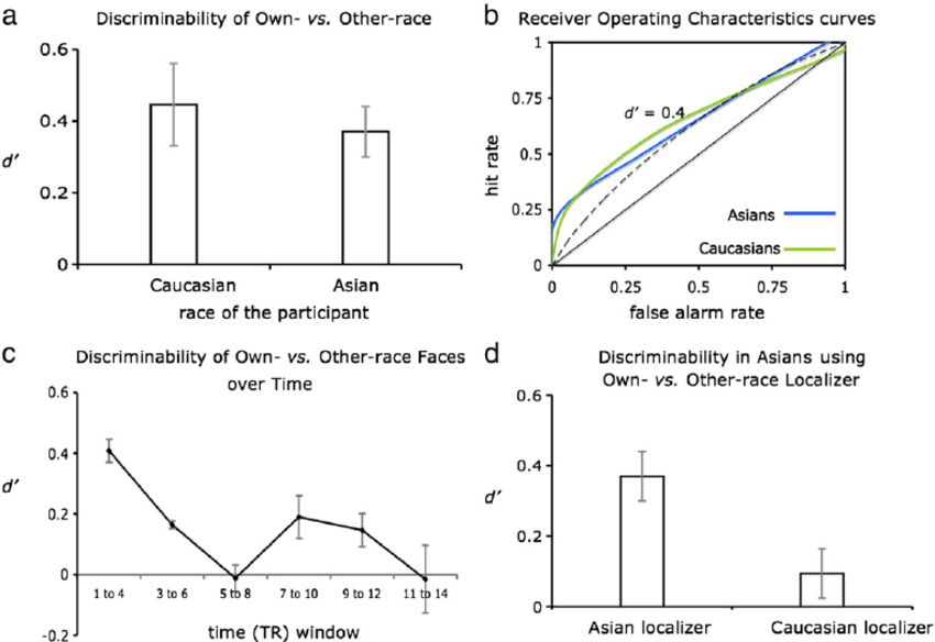
(Image from: Natu et al., 2010, NeuroImage).

Subscribe

Sign up with your email address to receive news and updates.

Thank you!
Back to Top
New Page

Powered by Squarespace